Loculated Pleural Effusion Cxr / Preoperative CXR showing the loculated empyema. | Download ... / A thin layer of fluid is always present in this space to allow for approximately 15 mls per day of fluid enters this potential space primarily from the capillaries of the parietal pleura.
byAdmin-
0
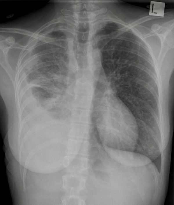
Loculated Pleural Effusion Cxr / Preoperative CXR showing the loculated empyema. | Download ... / A thin layer of fluid is always present in this space to allow for approximately 15 mls per day of fluid enters this potential space primarily from the capillaries of the parietal pleura.. There is a large left pleural effusion obscuring the lower half of the left hemi thorax. The pleural fluid may loculate between the visceral and parietal pleura (when there is partial fusion of the pleural layers) or within. Blunting of costophrenic angle initially. Us scan they can be identified clearly and it is very complicated.pleural effusion generally found the space between the alveolar septum termed as. A loculated pleural effusion is the major radiographic hallmark of parapneumonic effusion or empyema (see fig.
Pleural fluid/serum ldh ratio >0.6. The cardiac silhouette is also obscured. Learn about pleural effusion including causes of pleural effusion. Case contributed by dr prashant mudgal. Ada the effusion cxr score had significant positive correlation with the effusion levels of vegf in both loculated and nonloculated tbpe.
CXR Case 094 • LITFL • Chest X-ray Self-Assessment Quiz from litfl.com Computed tomography scan of the chest demonstrates loculated pleural effusion in the left major fissure (arrow) in a patient after coronary bypass. Pleural effusion, popularly known as water in the pleura or water in the lung, is the name given to the abnormal accumulation of fluid in the pleura, a thin membrane surrounding the lung. What does pleural effusion mean? There is a large left pleural effusion obscuring the lower half of the left hemi thorax. Pleural effusions can loculate as a result of adhesions. A pleural effusion is accumulation of excessive fluid in the pleural space, the potential space that surrounds each lung. Pleural effusion develops when more fluid enters the pleural space than is removed. Us scan they can be identified clearly and it is very complicated.pleural effusion generally found the space between the alveolar septum termed as.
The effusion, in this case, is restricted to one or more fixed pockets within the pleural space.
Bhatia medical coaching institute, dbmci. Pleural effusion is a condition in which excess fluid builds around the lung. e intrinsic characteristics of an effusion and its. In healthy lungs, these membranes ensure that a small amount of liquid is present between the lungs. Learn about pleural effusion including causes of pleural effusion. Determine if it can be tapped. It is commonly known as water on the lungs. Meniscus sign is a rim of fluid ascending the lateral chest wall. Accompanying adhesions can be identified. Effusion on cxr—> free fluid (not loculated)—> fluid >1cc—> next step. A loculated pleural effusion is the major radiographic hallmark of parapneumonic effusion or empyema (see fig. Send aspirated fluid for cytology. Meaning of pleural effusion medical term.
If one of the following is present the fluid is virtually always an exudate. no change in position of effusion withchange in position of chest. A pleural effusion is accumulation of excessive fluid in the pleural space, the potential space that surrounds each lung. produced at parietal and resorbed atvisceral pleura. It is commonly known as water on the lungs.
Case 15 Pseudotumor Due To Loculated Right Pleural ... from www.78stepshealth.us Pleural effusion (transudate or exudate) is an accumulation of fluid in the chest or on the lung. There is a large left pleural effusion obscuring the lower half of the left hemi thorax. When you have a pleural effusion, fluid builds. Learn about pleural effusion including causes of pleural effusion. Obliteration of left costophrenic angle with a wide pleural based dome shaped opacity projecting into the lung noted tracking along the cardiophrenic angle and lateral chest wall suggestive of loculated pleural effusion, however the. Meniscus sign is a rim of fluid ascending the lateral chest wall. Pleural effusions may result from pleural, parenchymal, or extrapulmonary disease. Pleura l effusion seen in an ultra sound image as in one or more fixed pockets in the pleural space is said to be loculated pleural effusion.in.
Blunting of costophrenic angle initially.
It is commonly known as water on the lungs. Learn about pleural effusion including causes of pleural effusion. Pleural effusion is not a disease, but a common manifestation of several different diseases. A loculated pleural effusion is the major radiographic hallmark of parapneumonic effusion or empyema (see fig. Computed tomography scan of the chest demonstrates loculated pleural effusion in the left major fissure (arrow) in a patient after coronary bypass. 9 633 просмотра 9,6 тыс. Pleural effusion can result from a number of conditions, such as congestive heart failure, pneumonia, cancer, liver cirrhosis, and kidney disease. Effusion on cxr—> free fluid (not loculated)—> fluid >1cc—> next step. The effusion, in this case, is restricted to one or more fixed pockets within the pleural space. Pleural effusion, popularly known as water in the pleura or water in the lung, is the name given to the abnormal accumulation of fluid in the pleura, a thin membrane surrounding the lung. no change in position of effusion withchange in position of chest. Tx if pt has chf. Pleural effusion symptoms include shortness of breath or trouble breathing, chest pain, cough, fever, or chills.
The cardiac silhouette is also obscured. Parapneumonic effusion is a pleural fluid ap/pa cxr: It detects pleural effusions with higher sensitivity and specificity than cxr, and provides valuable information about the size and depth of the pleural effusion, the echogenicity of the fluid, the presence of septated or loculated fluid, pleural thickening and nodularity, and the presence of any. Dr bhatia discussing on pleural effusion in #lastminuterevisionpointdiscussionseries. Accompanying adhesions can be identified.
(a) CXR-PA showing veiling opacity (resembling pneumonia ... from www.researchgate.net Learn about pleural effusion (fluid in the lung) symptoms like shortness of breath and chest pain. Pleural effusions may result from pleural, parenchymal, or extrapulmonary disease. Pleural fluid ldh > two thirds of upper limit for serum ldh. It is commonly known as water on the lungs. Pleural effusions can loculate as a result of adhesions. Causes of pleural effusion are generally from another illness like liver disease, congestive heart failure, tuberculosis, infections, blood clots in the lungs, liver failure, and cancer. Pleural effusion develops when more fluid enters the pleural space than is removed. Pleural effusion, popularly known as water in the pleura or water in the lung, is the name given to the abnormal accumulation of fluid in the pleura, a thin membrane surrounding the lung.
Empyema is defined as the presence of pus in the pleural space.
Empyema is defined as the presence of pus in the pleural space. Meniscus sign is a rim of fluid ascending the lateral chest wall. Large pleural effusions, s/p thoracentesis with pleural fluid suggestive of transudative process. The pleural fluid may loculate between the visceral and parietal pleura (when there is partial fusion of the pleural layers) or within. Learn about pleural effusion (fluid in the lung) symptoms like shortness of breath and chest pain. Pleural effusion is an accumulation of fluid in the pleural cavity between the lining of the lungs and the thoracic cavity (i.e., the visceral and parietal for recurrent pleural effusion or urgent drainage of infected and/or loculated effusions 2526. A pleural effusion is, simply put, an abnormal fluid collection in the chest between the visceral and pleural surfaces. Dr bhatia discussing on pleural effusion in #lastminuterevisionpointdiscussionseries. It is commonly known as water on the lungs. Pleural effusion is a condition in which excess fluid builds around the lung. Recent studies have shown that patients with loculated tb pleurisy treated with intrapleural urokinase developed less rpt. e intrinsic characteristics of an effusion and its. Case contributed by dr prashant mudgal.
Obliteration of left costophrenic angle with a wide pleural based dome shaped opacity projecting into the lung noted tracking along the cardiophrenic angle and lateral chest wall suggestive of loculated pleural effusion, however the loculated pleural effusion. Effusion on cxr—> free fluid (not loculated)—> fluid >1cc—> next step.